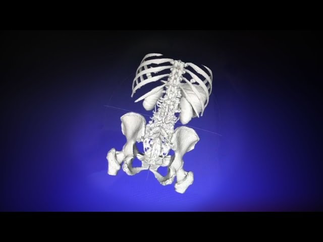
With advances in computing and software power, physicians can now create virtual 3-dimensional models of patient anatomy using data from 2-dimensional computed tomography imaging. With augmented reality, users can interact with each other and the model, even over long distances, for purposes of teaching or preprocedural decision-making. This video illustrates an example of the process; the 3-dimensional model has been loaded onto a HoloLens (Microsoft) augmented reality viewing device for inspection and manipulation. By performing an air tap gesture similar to a mouse click, the user can disassemble components of the 3-dimensional model to understand the anatomic relations between each. For more detail, see https://ja.ma/2KCTw7q.